Παρασκευή 4 Μαρτίου 2016

Cannabis use in psychotic patients is linked to worse outcomes

Cannabis use among patients with first episode psychosis is associated with substantially worse clinical outcomes, a large study published in BMJ Open has found.1Psychotic patients with a history of...
recent?d=yIl2AUoC8zA recent?d=dnMXMwOfBR0 recent?i=eEv2uV84iEg:nA_NiJAAMOg:V_sGLiP recent?d=qj6IDK7rITs recent?i=eEv2uV84iEg:nA_NiJAAMOg:gIN9vFw recent?d=l6gmwiTKsz0 recent?d=7Q72WNTAKBA recent?i=eEv2uV84iEg:nA_NiJAAMOg:F7zBnMy recent?i=eEv2uV84iEg:nA_NiJAAMOg:-BTjWOF


from #AlexandrosSfakianakis via Alexandros G.Sfakianakis on Inoreader http://ift.tt/21b31ud
via IFTTT

Long term aspirin may reduce overall cancer risk

Taking low dose aspirin regularly for at least six years is associated with a modest but significant reduction in the risk of cancer, especially cancer of the gastrointestinal tract, research...
recent?d=yIl2AUoC8zA recent?d=dnMXMwOfBR0 recent?i=9pvrpVg1C9I:lUcqyXQQTSw:V_sGLiP recent?d=qj6IDK7rITs recent?i=9pvrpVg1C9I:lUcqyXQQTSw:gIN9vFw recent?d=l6gmwiTKsz0 recent?d=7Q72WNTAKBA recent?i=9pvrpVg1C9I:lUcqyXQQTSw:F7zBnMy recent?i=9pvrpVg1C9I:lUcqyXQQTSw:-BTjWOF


from #AlexandrosSfakianakis via Alexandros G.Sfakianakis on Inoreader http://ift.tt/21J1YXM
via IFTTT

IJERPH, Vol. 13, Pages 281: Toxicity of Smokeless Tobacco Extract after 184-Day Repeated Oral Administration in Rats

The use of smokeless tobacco (ST) is growing rapidly and globally. The consumption of ST is associated with an increased risk for developing chronic diseases, such as diabetes, hypercholesterolemia, and myocardial infarction, and has led to many public health problems. It is very important to access the toxicity of ST. This experiment presents data from 184-day toxicology studies in Sprague-Dawley (SD) rats designed to characterize the chronic effects of a smokeless tobacco extract (STE). The control group and treatment groups were matched for a range of nicotine levels. Animals were given STE by oral gavage with doses of 3.75 (low-dose), 7.50 (mid-dose) and 15.00 (high-dose) mg·nicotine/kg body weight/day for 184 days, followed by 30 days for recovery. Variables evaluated included body weights, feed consumption, clinical observations, clinical and anatomic pathology (including organ weights), and histopathology. Decreased body weights and organ weights (heart, liver and kidney) were found in animals in the mid-dose and high-dose groups. STE also showed moderate and reversible toxicity in esophagus, stomach, liver, kidney and lung.

from #AlexandrosSfakianakis via Alexandros G.Sfakianakis on Inoreader http://ift.tt/1WYM4Sl
via IFTTT

In-vivo evaluation of apocynin for prevention of Helicobacter pylori-induced gastric carcinogenesis.

The emergence of antibiotic-resistant Helicobacter pylori strains impacts the efficacy of eradication therapy and promotes the development of alternative treatment strategies. Apocynin inhibits neutrophil NADPH oxidase and hence may decrease reactive oxygen species-mediated tissue damage in H. pylori-infected stomach tissue. Apocynin was tested in vitro for its cytotoxic and direct antibacterial effects. The therapeutic efficacy of orally administered apocynin (100 mg/kg/day through drinking water or 200 mg/kg/day through combined administration of drinking water and slow-release formulation) was assessed at 9 weeks after infection in the Mongolian gerbil model. Bacterial burdens were quantified by viable plate count and quantitative PCR. Histopathological evaluation of antrum and pylorus provided insight into mucosal inflammation and injury. Apocynin showed no cytotoxic or direct antibacterial effects in vitro or in vivo. Nine weeks of apocynin treatment at 200 mg/kg/day reduced active H. pylori gastritis as neutrophil infiltration in the mucous neck region and pit abscess formation decreased significantly. In our gerbil model, prolonged high-dose apocynin treatment significantly improved H. pylori-induced pit abscess formation without indications of drug toxicity and thus further investigation of the dosage regimen and formulation and the long-term impact on neoplastic development should be carried out. Copyright (C) 2016 Wolters Kluwer Health, Inc. All rights reserved.

from #AlexandrosSfakianakis via Alexandros G.Sfakianakis on Inoreader http://ift.tt/1VUJOLK
via IFTTT

Determination of the binding properties of the uremic toxin phenylacetic acid to Human Serum Albumin

Publication date: Available online 3 March 2016
Source:Biochimie
Author(s): Juliana F. Saldanha, Dan Yi, Milena B. Stockler-Pinto, Hédi A. Soula, Stéphane Chambert, Denis Fouque, Denise Mafra, Christophe O. Soulage
Uremic toxins are compounds normally excreted in urine that accumulate in patients with chronic kidney disease as a result of decreased renal clearance. Phenylacetic acid (PAA) has been identified as a new protein bound uremic toxin. The purpose of this study was to investigate in vitro the interaction between PAA and human serum albumin (HSA) at physiological and pathological concentrations. We used ultrafiltration to show that there is a single high-affinity binding site for PAA on HSA, with a binding constant on the order of 3.4×10⁴ M-1 and a maximal stoichiometry of 1.61 mole per mole. The PAA, at the concentration reported in end-stage renal patients, was 26% bound to albumin. Fluorescent probe competition experiments demonstrated that PAA did not bind to Sudlow’s site I (in subdomain IIA) and only weakly bind to Sudlow‘s site II (in subdomain IIIA). The PAA showed no competition with other protein-bound uremic toxins such as p-cresyl-sulfate or indoxyl sulphate for binding to serum albumin. Our results provide evidence that human serum albumin can act as carrier protein for phenylacetic acid.


from #AlexandrosSfakianakis via Alexandros G.Sfakianakis on Inoreader http://ift.tt/1nkFZn4
via IFTTT

Loss of a Single Mcl-1 Allele Inhibits MYC-Driven Lymphomagenesis by Sensitizing Pro-B Cells to Apoptosis

Publication date: Available online 3 March 2016
Source:Cell Reports
Author(s): Stephanie Grabow, Alex R.D. Delbridge, Brandon J. Aubrey, Cassandra J. Vandenberg, Andreas Strasser
MCL-1 is critical for progenitor cell survival during emergency hematopoiesis, but its role in sustaining cells undergoing transformation and in lymphomagenesis is only poorly understood. We investigated the importance of MCL-1 in the survival of B lymphoid progenitors undergoing MYC-driven transformation and its functional interactions with pro-apoptotic BIM and PUMA and the tumor suppressor p53 in lymphoma development. Loss of one Mcl-1 allele almost abrogated MYC-driven-lymphoma development owing to a reduction in lymphoma initiating pre-B cells. Although loss of the p53 target PUMA had minor impact, loss of one p53 allele substantially accelerated lymphoma development when MCL-1 was limiting, most likely because p53 loss also causes defects in non-apoptotic tumor suppressive processes. Remarkably, loss of BIM restored the survival of lymphoma initiating cells and rate of tumor development. Thus, MCL-1 has a major role in lymphoma initiating pro-B cells to oppose BIM, which is upregulated in response to oncogenic stress.

Graphical abstract

image

Teaser

MCL-1 is overexpressed in various human cancers. Grabow et al. reveal the importance of MCL-1 for the survival of B cell progenitors undergoing neoplastic transformation and in lymphomagenesis. Given that non-transformed cells appear to be less dependent on MCL-1 than malignant ones, inhibitors of MCL-1 may be useful in cancer therapy.


from #AlexandrosSfakianakis via Alexandros G.Sfakianakis on Inoreader http://ift.tt/1UDW2Kc
via IFTTT

The P72R Polymorphism of p53 Predisposes to Obesity and Metabolic Dysfunction

Publication date: Available online 3 March 2016
Source:Cell Reports
Author(s): Che-Pei Kung, Julia I-Ju Leu, Subhasree Basu, Sakina Khaku, Frederick Anokye-Danso, Qin Liu, Donna L. George, Rexford S. Ahima, Maureen E. Murphy
p53 is well known for its tumor suppressor role, but this protein also has a poorly understood role in the regulation of metabolism. Human studies have implicated a common polymorphism at codon 72 of p53 in diabetic and pre-diabetic phenotypes. To understand this role, we utilized a humanized mouse model of the p53 codon 72 variants and monitored these mice following challenge with a high-fat diet (HFD). Mice with the arginine 72 (R72) variant of p53 developed more-severe obesity and glucose intolerance on a HFD, compared to mice with the proline 72 variant (P72). R72 mice developed insulin resistance, islet hypertrophy, increased infiltration of immune cells, and fatty liver disease. Gene expression analyses and studies with small-molecule inhibitors indicate that the p53 target genes Tnf and Npc1l1 underlie this phenotype. These results shed light on the role of p53 in obesity, metabolism, and inflammation.

Graphical abstract

image

Teaser

Kung et al. show that the R72 variant of p53 leads to increased obesity and glucose intolerance in mice fed a high-fat diet. They identify two p53 target genes, Tnf and Npc1l1, that are preferentially bound by R72 and that are responsible for this phenotype.


from #AlexandrosSfakianakis via Alexandros G.Sfakianakis on Inoreader http://ift.tt/1UDW2tY
via IFTTT

Dual and Opposing Roles of MicroRNA-124 in Epilepsy Are Mediated through Inflammatory and NRSF-Dependent Gene Networks

Publication date: Available online 3 March 2016
Source:Cell Reports
Author(s): Gary P. Brennan, Deblina Dey, Yuncai Chen, Katelin P. Patterson, Eric J. Magnetta, Alicia M. Hall, Celine M. Dube, Yu-Tang Mei, Tallie Z. Baram
Insult-provoked transformation of neuronal networks into epileptic ones involves multiple mechanisms. Intervention studies have identified both dysregulated inflammatory pathways and NRSF-mediated repression of crucial neuronal genes as contributors to epileptogenesis. However, it remains unclear how epilepsy-provoking insults (e.g., prolonged seizures) induce both inflammation and NRSF and whether common mechanisms exist. We examined miR-124 as a candidate dual regulator of NRSF and inflammatory pathways. Status epilepticus (SE) led to reduced miR-124 expression via SIRT1—and, in turn, miR-124 repression—via C/EBPα upregulated NRSF. We tested whether augmenting miR-124 after SE would abort epileptogenesis by preventing inflammation and NRSF upregulation. SE-sustaining animals developed epilepsy, but supplementing miR-124 did not modify epileptogenesis. Examining this result further, we found that synthetic miR-124 not only effectively blocked NRSF upregulation and rescued NRSF target genes, but also augmented microglia activation and inflammatory cytokines. Thus, miR-124 attenuates epileptogenesis via NRSF while promoting epilepsy via inflammation.

Graphical abstract

image

Teaser

Brennan et al. find that miR-124 is involved in insult-induced epilepsy. They find that activation of SIRT1 represses miR124. This repression affects miR-124 targeting of NRSF; however, it may also act to reduce inflammation that contributes to epilepsy development. Thus, miR-124 has opposing roles in the development of epilepsy.


from #AlexandrosSfakianakis via Alexandros G.Sfakianakis on Inoreader http://ift.tt/1UDW2tW
via IFTTT

Localized Translation of gurken/TGF-α mRNA during Axis Specification Is Controlled by Access to Orb/CPEB on Processing Bodies

Publication date: Available online 3 March 2016
Source:Cell Reports
Author(s): Alexander Davidson, Richard M. Parton, Catherine Rabouille, Timothy T. Weil, Ilan Davis
In Drosophila oocytes, gurken/TGF-α mRNA is essential for establishing the future embryonic axes. gurken remains translationally silent during transport from its point of synthesis in nurse cells to its final destination in the oocyte, where it associates with the edge of processing bodies. Here we show that, in nurse cells, gurken is kept translationally silent by the lack of sufficient Orb/CPEB, its translational activator. Processing bodies in nurse cells have a similar protein complement and ultrastructure to those in the oocyte, but they markedly less Orb and do not associate with gurken mRNA. Ectopic expression of Orb in nurse cells at levels similar to the wild-type oocyte dorso-anterior corner at mid-oogenesis is sufficient to cause gurken mRNA to associate with processing bodies and translate prematurely. We propose that controlling the spatial distribution of translational activators is a fundamental mechanism for regulating localized translation.

Graphical abstract

image

Teaser

Localized transcripts that determine polarity are thought to be silenced during their transport by binding to translational repressors. Davidson et al. show that gurken mRNA, which sets up the primary Drosophila body axes, is instead regulated through lack of a translational activator, Orb, during its transport.


from #AlexandrosSfakianakis via Alexandros G.Sfakianakis on Inoreader http://ift.tt/1UDW2tR
via IFTTT

Fibroblast Growth Factor 21 Mediates Glycemic Regulation by Hepatic JNK

Publication date: Available online 3 March 2016
Source:Cell Reports
Author(s): Santiago Vernia, Julie Cavanagh-Kyros, Tamera Barrett, Cathy Tournier, Roger J. Davis
The cJun NH2-terminal kinase (JNK)-signaling pathway is implicated in metabolic syndrome, including dysregulated blood glucose concentration and insulin resistance. Fibroblast growth factor 21 (FGF21) is a target of the hepatic JNK-signaling pathway and may contribute to the regulation of glycemia. To test the role of FGF21, we established mice with selective ablation of the Fgf21 gene in hepatocytes. FGF21 deficiency in the liver caused marked loss of FGF21 protein circulating in the blood. Moreover, the protective effects of hepatic JNK deficiency to suppress metabolic syndrome in high-fat diet-fed mice were not observed in mice with hepatocyte-specific FGF21 deficiency, including reduced blood glucose concentration and reduced intolerance to glucose and insulin. Furthermore, we show that JNK contributes to the regulation of hepatic FGF21 expression during fasting/feeding cycles. These data demonstrate that the hepatokine FGF21 is a key mediator of JNK-regulated metabolic syndrome.

Graphical abstract

image

Teaser

Vernia et al. examine the role of circulating FGF21 in the response to JNK signaling. Hepatic JNK deficiency promotes expression of FGF21 and improves glycemia. Liver-specific ablation of the Fgf21 gene prevents this improvement of glycemia. These data argue that FGF21 mediates metabolic actions of hepatic JNK.


from #AlexandrosSfakianakis via Alexandros G.Sfakianakis on Inoreader http://ift.tt/1UDW0C8
via IFTTT

Landscape and Dynamics of Transcription Initiation in the Malaria Parasite Plasmodium falciparum

Publication date: Available online 3 March 2016
Source:Cell Reports
Author(s): Sophie H. Adjalley, Christophe D. Chabbert, Bernd Klaus, Vicent Pelechano, Lars M. Steinmetz
A comprehensive map of transcription start sites (TSSs) across the highly AT-rich genome of P. falciparum would aid progress toward deciphering the molecular mechanisms that underlie the timely regulation of gene expression in this malaria parasite. Using high-throughput sequencing technologies, we generated a comprehensive atlas of transcription initiation events at single-nucleotide resolution during the parasite intra-erythrocytic developmental cycle. This detailed analysis of TSS usage enabled us to define architectural features of plasmodial promoters. We demonstrate that TSS selection and strength are constrained by local nucleotide composition. Furthermore, we provide evidence for coordinate and stage-specific TSS usage from distinct sites within the same transcription unit, thereby producing transcript isoforms, a subset of which are developmentally regulated. This work offers a framework for further investigations into the interactions between genomic sequences and regulatory factors governing the complex transcriptional program of this major human pathogen.

Graphical abstract

image

Teaser

The genome of P. falciparum is characterized by an extremely AT-biased nucleotide composition. Adjalley et al. conducted a survey of transcription initiation events throughout the malaria parasite’s blood cycle. They detect coordinate and stage-specific transcription initiation and extract sequence and chromatin signatures from promoter regions.


from #AlexandrosSfakianakis via Alexandros G.Sfakianakis on Inoreader http://ift.tt/1UDW0lQ
via IFTTT

Plasticity between Epithelial and Mesenchymal States Unlinks EMT from Metastasis-Enhancing Stem Cell Capacity

Publication date: Available online 3 March 2016
Source:Cell Reports
Author(s): Evelyne Beerling, Daniëlle Seinstra, Elzo de Wit, Lennart Kester, Daphne van der Velden, Carrie Maynard, Ronny Schäfer, Paul van Diest, Emile Voest, Alexander van Oudenaarden, Nienke Vrisekoop, Jacco van Rheenen
Forced overexpression and/or downregulation of proteins regulating epithelial-to-mesenchymal transition (EMT) has been reported to alter metastasis by changing migration and stem cell capacity of tumor cells. However, these manipulations artificially keep cells in fixed states, while in vivo cells may adapt transient and reversible states. Here, we have tested the existence and role of epithelial-mesenchymal plasticity in metastasis of mammary tumors without artificially modifying EMT regulators. In these tumors, we found by intravital microscopy that the motile tumor cells have undergone EMT, while their epithelial counterparts were not migratory. Moreover, we found that epithelial-mesenchymal plasticity renders any EMT-induced stemness differences, as reported previously, irrelevant for metastatic outgrowth, because mesenchymal cells that arrive at secondary sites convert to the epithelial state within one or two divisions, thereby obtaining the same stem cell potential as their arrived epithelial counterparts. We conclude that epithelial-mesenchymal plasticity supports migration but additionally eliminates stemness-enhanced metastatic outgrowth differences.

Graphical abstract

image

Teaser

Beerling et al. identified a previously undetectable pool of cells in epithelial breast tumors that have undergone EMT without experimental induction. These cells are motile when disseminating and revert back to the epithelial state upon metastatic outgrowth. This epithelial-mesenchymal plasticity equalizes metastatic outgrowth potential between epithelial and mesenchymal tumor cells.


from #AlexandrosSfakianakis via Alexandros G.Sfakianakis on Inoreader http://ift.tt/1UDW2tG
via IFTTT

YAP Induces Human Naive Pluripotency

Publication date: Available online 3 March 2016
Source:Cell Reports
Author(s): Han Qin, Miroslav Hejna, Yanxia Liu, Michelle Percharde, Mark Wossidlo, Laure Blouin, Jens Durruthy-Durruthy, Priscilla Wong, Zhongxia Qi, Jingwei Yu, Lei S. Qi, Vittorio Sebastiano, Jun S. Song, Miguel Ramalho-Santos
The human naive pluripotent stem cell (PSC) state, corresponding to a pre-implantation stage of development, has been difficult to capture and sustain in vitro. We report that the Hippo pathway effector YAP is nuclearly localized in the inner cell mass of human blastocysts. Overexpression of YAP in human embryonic stem cells (ESCs) and induced PSCs (iPSCs) promotes the generation of naive PSCs. Lysophosphatidic acid (LPA) can partially substitute for YAP to generate transgene-free human naive PSCs. YAP- or LPA-induced naive PSCs have a rapid clonal growth rate, a normal karyotype, the ability to form teratomas, transcriptional similarities to human pre-implantation embryos, reduced heterochromatin levels, and other hallmarks of the naive state. YAP/LPA act in part by suppressing differentiation-inducing effects of GSK3 inhibition. CRISPR/Cas9-generated YAP−/− cells have an impaired ability to form colonies in naive but not primed conditions. These results uncover an unexpected role for YAP in the human naive state, with implications for early human embryology.

Graphical abstract

image

Teaser

Qin et al. find that the Hippo pathway effector YAP induces and sustains the naive state of human pluripotency, which corresponds to preimplantation cells. Lysophosphatidic acid, a small molecule that activates YAP, has a similar effect, opening up opportunities for studies of human embryology and disease modeling.


from #AlexandrosSfakianakis via Alexandros G.Sfakianakis on Inoreader http://ift.tt/1UDW2do
via IFTTT

Sialic Acid-Binding Immunoglobulin-like Lectin G Promotes Atherosclerosis and Liver Inflammation by Suppressing the Protective Functions of B-1 Cells

Publication date: Available online 3 March 2016
Source:Cell Reports
Author(s): Sabrina Gruber, Tim Hendrikx, Dimitrios Tsiantoulas, Maria Ozsvar-Kozma, Laura Göderle, Ziad Mallat, Joseph L. Witztum, Ronit Shiri-Sverdlov, Lars Nitschke, Christoph J. Binder
Atherosclerosis is initiated and sustained by hypercholesterolemia, which results in the generation of oxidized LDL (OxLDL) and other metabolic byproducts that trigger inflammation. Specific immune responses have been shown to modulate the inflammatory response during atherogenesis. The sialic acid-binding immunoglobulin-like lectin G (Siglec-G) is a negative regulator of the functions of several immune cells, including myeloid cells and B-1 cells. Here, we show that deficiency of Siglec-G in atherosclerosis-prone mice inhibits plaque formation and diet-induced hepatic inflammation. We further demonstrate that selective deficiency of Siglec-G in B cells alone is sufficient to mediate these effects. Levels of B-1 cell-derived natural IgM with specificity for OxLDL were significantly increased in the plasma and peritoneal cavity of Siglec-G-deficient mice. Consistent with the neutralizing functions of OxLDL-specific IgM, Siglec-G-deficient mice were protected from OxLDL-induced sterile inflammation. Thus, Siglec-G promotes atherosclerosis and hepatic inflammation by suppressing protective anti-inflammatory effector functions of B cells.

Graphical abstract

image

Teaser

Gruber et al. demonstrate that Siglec-G deficiency protects from oxidized LDL-induced inflammation through the expansion of B-1 cells secreting natural IgM antibodies, leading to reduced atherosclerosis and hepatic inflammation. CXCL1 represents a common pro-inflammatory factor that is reduced as a result of Siglec-G deficiency.


from #AlexandrosSfakianakis via Alexandros G.Sfakianakis on Inoreader http://ift.tt/1UDW0lE
via IFTTT

Central Insulin Action Activates Kupffer Cells by Suppressing Hepatic Vagal Activation via the Nicotinic Alpha 7 Acetylcholine Receptor

Publication date: Available online 3 March 2016
Source:Cell Reports
Author(s): Kumi Kimura, Mamoru Tanida, Naoto Nagata, Yuka Inaba, Hitoshi Watanabe, Mayumi Nagashimada, Tsuguhito Ota, Shun-ichiro Asahara, Yoshiaki Kido, Michihiro Matsumoto, Koji Toshinai, Masamitsu Nakazato, Toshishige Shibamoto, Shuichi Kaneko, Masato Kasuga, Hiroshi Inoue
Central insulin action activates hepatic IL-6/STAT3 signaling, which suppresses the gene expression of hepatic gluconeogenic enzymes. The vagus nerve plays an important role in this centrally mediated hepatic response; however, the precise mechanism underlying this brain-liver interaction is unclear. Here, we present our findings that the vagus nerve suppresses hepatic IL-6/STAT3 signaling via α7-nicotinic acetylcholine receptors (α7-nAchR) on Kupffer cells, and that central insulin action activates hepatic IL-6/STAT3 signaling by suppressing vagal activity. Indeed, central insulin-mediated hepatic IL-6/STAT3 activation and gluconeogenic gene suppression were impeded in mice with hepatic vagotomy, pharmacological cholinergic blockade, or α7-nAchR deficiency. In high-fat diet-induced obese and insulin-resistant mice, control of the vagus nerve by central insulin action was disturbed, inducing a persistent increase of inflammatory cytokines. These findings suggest that dysregulation of the α7-nAchR-mediated control of Kupffer cells by central insulin action may affect the pathogenesis of chronic hepatic inflammation in obesity.

Graphical abstract

image

Teaser

In this paper, Kimura et al. show a mechanism of the central insulin-mediated hepatic response, where central insulin action—known to suppress hepatic glucose production via hepatic IL-6/STAT3 activation—mitigates the α7-nAchR-dependent downregulation of IL-6 expression in Kupffer cells by the vagus nerve.


from #AlexandrosSfakianakis via Alexandros G.Sfakianakis on Inoreader http://ift.tt/1UDW2df
via IFTTT

Global Analysis of Cellular Protein Flux Quantifies the Selectivity of Basal Autophagy

Publication date: Available online 3 March 2016
Source:Cell Reports
Author(s): Tian Zhang, Shichen Shen, Jun Qu, Sina Ghaemmaghami
In eukaryotic cells, macroautophagy is a catabolic pathway implicated in the degradation of long-lived proteins and damaged organelles. Although it has been demonstrated that macroautophagy can selectively degrade specific targets, its contribution to the basal turnover of cellular proteins has not been quantified on proteome-wide scales. In this study, we created autophagy-deficient primary human fibroblasts and quantified the resulting changes in basal degradative flux by dynamic proteomics. Our results provide a global comparison of protein half-lives between wild-type and autophagy-deficient cells. The data indicate that in quiescent fibroblasts, macroautophagy contributes to the basal turnover of a substantial fraction of the proteome at varying levels. As contrasting examples, we demonstrate that the proteasome and CCT/TRiC chaperonin are robust substrates of basal autophagy, whereas the ribosome is largely protected under basal conditions. This selectivity may establish a proteostatic feedback mechanism that stabilizes the proteasome and CCT/TRiC when autophagy is inhibited.

Graphical abstract

image

Teaser

Macroautophagy is a catabolic pathway for the degradation of proteins in eukaryotic cells. Zhang et al. quantified the relative contribution of macroautophagy to basal proteome turnover by comparing protein half-lives between wild-type and autophagy-deficient fibroblasts. The data provide a global map of the selectivity of macroautophagy in human cells.


from #AlexandrosSfakianakis via Alexandros G.Sfakianakis on Inoreader http://ift.tt/1UDW0lz
via IFTTT

Context-Dependent Development of Lymphoid Stroma from Adult CD34+ Adventitial Progenitors

Publication date: Available online 3 March 2016
Source:Cell Reports
Author(s): Katarzyna M. Sitnik, Kerstin Wendland, Holger Weishaupt, Heli Uronen-Hansson, Andrea J. White, Graham Anderson, Knut Kotarsky, William W. Agace
Despite the key role of primary and secondary lymphoid organ stroma in immunity, our understanding of the heterogeneity and ontogeny of these cells remains limited. Here, we identify a functionally distinct subset of BP3PDPN+PDGFRβ++CD34+ stromal adventitial cells in both lymph nodes (LNs) and thymus that is located within the vascular niche surrounding PDPNPDGFRβ+Esam-1+ITGA7+ pericytes. CD34+ adventitial cells developed in late embryonic thymus and in postnatal LNs and in the thymus originated, along with pericytes, from a common anlage-seeding progenitor population. Using lymphoid organ re-aggregate grafts, we demonstrate that adult CD34+ adventitial cells are capable of differentiating into multiple lymphoid stroma-like subsets including pericyte-, FRC-, MRC-, and FDC-like cells, the development of which was lymphoid environment-dependent. These findings extend the current understanding of lymphoid mesenchymal cell heterogeneity and highlight a role of the CD34+ adventitia as a potential ubiquitous source of lymphoid stromal precursors in postnatal tissues.

Graphical abstract

image

Teaser

Sitnik et al. comparatively assess the stromal cell composition of the thymus and lymph nodes. The authors identify CD34+ adventitial cells as a conserved component of the thymic and lymph node vascular niche and demonstrate that this subset contains adult progenitors with lymphoid stroma-like subset potential in vivo.


from #AlexandrosSfakianakis via Alexandros G.Sfakianakis on Inoreader http://ift.tt/1UDW2db
via IFTTT

Large-Scale Profiling of Kinase Dependencies in Cancer Cell Lines

Publication date: Available online 3 March 2016
Source:Cell Reports
Author(s): James Campbell, Colm J. Ryan, Rachel Brough, Ilirjana Bajrami, Helen N. Pemberton, Irene Y. Chong, Sara Costa-Cabral, Jessica Frankum, Aditi Gulati, Harriet Holme, Rowan Miller, Sophie Postel-Vinay, Rumana Rafiq, Wenbin Wei, Chris T. Williamson, David A. Quigley, Joe Tym, Bissan Al-Lazikani, Timothy Fenton, Rachael Natrajan, Sandra J. Strauss, Alan Ashworth, Christopher J. Lord
One approach to identifying cancer-specific vulnerabilities and therapeutic targets is to profile genetic dependencies in cancer cell lines. Here, we describe data from a series of siRNA screens that identify the kinase genetic dependencies in 117 cancer cell lines from ten cancer types. By integrating the siRNA screen data with molecular profiling data, including exome sequencing data, we show how vulnerabilities/genetic dependencies that are associated with mutations in specific cancer driver genes can be identified. By integrating additional data sets into this analysis, including protein-protein interaction data, we also demonstrate that the genetic dependencies associated with many cancer driver genes form dense connections on functional interaction networks. We demonstrate the utility of this resource by using it to predict the drug sensitivity of genetically or histologically defined subsets of tumor cell lines, including an increased sensitivity of osteosarcoma cell lines to FGFR inhibitors and SMAD4 mutant tumor cells to mitotic inhibitors.

Graphical abstract

image

Teaser

Campbell et al. use parallel siRNA screens to identify the kinase dependencies of 117 cancer cell lines from ten cancer types. They use this resource to identify kinase dependencies associated with specific cancer types or driver genes and show that the integration of protein interaction networks facilitates the interpretation of these dependencies.


from #AlexandrosSfakianakis via Alexandros G.Sfakianakis on Inoreader http://ift.tt/1UDW05h
via IFTTT

Multilevel Genomics-Based Taxonomy of Renal Cell Carcinoma

Publication date: Available online 3 March 2016
Source:Cell Reports
Author(s): Fengju Chen, Yiqun Zhang, Yasin Şenbabaoğlu, Giovanni Ciriello, Lixing Yang, Ed Reznik, Brian Shuch, Goran Micevic, Guillermo De Velasco, Eve Shinbrot, Michael S. Noble, Yiling Lu, Kyle R. Covington, Liu Xi, Jennifer A. Drummond, Donna Muzny, Hyojin Kang, Junehawk Lee, Pheroze Tamboli, Victor Reuter, Carl Simon Shelley, Benny A. Kaipparettu, Donald P. Bottaro, Andrew K. Godwin, Richard A. Gibbs, Gad Getz, Raju Kucherlapati, Peter J. Park, Chris Sander, Elizabeth P. Henske, Jane H. Zhou, David J. Kwiatkowski, Thai H. Ho, Toni K. Choueiri, James J. Hsieh, Rehan Akbani, Gordon B. Mills, A. Ari Hakimi, David A. Wheeler, Chad J. Creighton
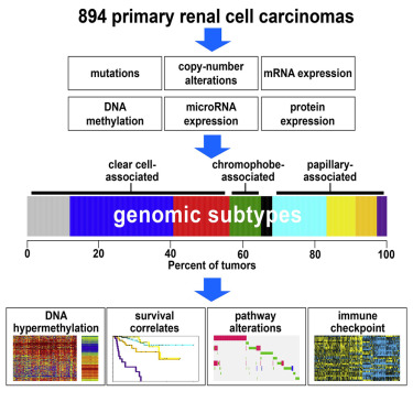
On the basis of multidimensional and comprehensive molecular characterization (including DNA methalylation and copy number, RNA, and protein expression), we classified 894 renal cell carcinomas (RCCs) of various histologic types into nine major genomic subtypes. Site of origin within the nephron was one major determinant in the classification, reflecting differences among clear cell, chromophobe, and papillary RCC. Widespread molecular changes associated with TFE3 gene fusion or chromatin modifier genes were present within a specific subtype and spanned multiple subtypes. Differences in patient survival and in alteration of specific pathways (including hypoxia, metabolism, MAP kinase, NRF2-ARE, Hippo, immune checkpoint, and PI3K/AKT/mTOR) could further distinguish the subtypes. Immune checkpoint markers and molecular signatures of T cell infiltrates were both highest in the subtype associated with aggressive clear cell RCC. Differences between the genomic subtypes suggest that therapeutic strategies could be tailored to each RCC disease subset.

Graphical abstract

image

Teaser

Chen et al. comprehensively analyze 894 renal cell carcinomas, incorporating data on DNA mutation and copy, DNA methylation, and gene expression. The cancers were thus classified into nine major subtypes, each one being distinct in terms of altered pathways and patient survival associations.


from #AlexandrosSfakianakis via Alexandros G.Sfakianakis on Inoreader http://ift.tt/1UDW1WV
via IFTTT

p73 Is Required for Multiciliogenesis and Regulates the Foxj1-Associated Gene Network

Publication date: Available online 3 March 2016
Source:Cell Reports
Author(s): Clayton B. Marshall, Deborah J. Mays, J. Scott Beeler, Jennifer M. Rosenbluth, Kelli L. Boyd, Gabriela L. Santos Guasch, Timothy M. Shaver, Lucy J. Tang, Qi Liu, Yu Shyr, Bryan J. Venters, Mark A. Magnuson, Jennifer A. Pietenpol
We report that p73 is expressed in multiciliated cells (MCCs), is required for MCC differentiation, and directly regulates transcriptional modulators of multiciliogenesis. Loss of ciliary biogenesis provides a unifying mechanism for many phenotypes observed in p73 knockout mice including hydrocephalus; hippocampal dysgenesis; sterility; and chronic inflammation/infection of lung, middle ear, and sinus. Through p73 and p63 ChIP-seq using murine tracheal cells, we identified over 100 putative p73 target genes that regulate MCC differentiation and homeostasis. We validated Foxj1, a transcriptional regulator of multiciliogenesis, and many other cilia-associated genes as direct target genes of p73 and p63. We show p73 and p63 are co-expressed in a subset of basal cells and suggest that p73 marks these cells for MCC differentiation. In summary, p73 is essential for MCC differentiation, functions as a critical regulator of a transcriptome required for MCC differentiation, and, like p63, has an essential role in development of tissues.

Graphical abstract

image

Teaser

Using a p73-deficient mouse model, Marshall et al. show that p73 is required for MCC differentiation. ChIP-seq of murine tracheal cells reveals many p73 target genes that regulate MCC differentiation. Lack of expression of key transcriptional regulators of ciliogenesis provides a mechanistic basis for the multiple defects in p73-deficient mice.


from #AlexandrosSfakianakis via Alexandros G.Sfakianakis on Inoreader http://ift.tt/1UDW1WT
via IFTTT

CARMA3 Is a Host Factor Regulating the Balance of Inflammatory and Antiviral Responses against Viral Infection

Publication date: Available online 3 March 2016
Source:Cell Reports
Author(s): Changying Jiang, Zhicheng Zhou, Yanping Quan, Shilei Zhang, Tingting Wang, Xueqiang Zhao, Clayton Morrison, Mark T. Heise, Wenqian He, Matthew S. Miller, Xin Lin
Host response to RNA virus infection is sensed by RNA sensors such as RIG-I, which induces MAVS-mediated NF-κB and IRF3 activation to promote inflammatory and antiviral responses, respectively. Here, we have found that CARMA3, a scaffold protein previously shown to mediate NF-κB activation induced by GPCR and EGFR, positively regulates MAVS-induced NF-κB activation. However, our data suggest that CARMA3 sequesters MAVS from forming high-molecular-weight aggregates, thereby suppressing TBK1/IRF3 activation. Interestingly, following NF-κB activation upon virus infection, CARMA3 is targeted for proteasome-dependent degradation, which releases MAVS to activate IRF3. When challenged with vesicular stomatitis virus or influenza A virus, CARMA3-deficient mice showed reduced disease symptoms compared to those of wild-type mice as a result of less inflammation and a stronger ability to clear infected virus. Altogether, our results reveal the role of CARMA3 in regulating the balance of host antiviral and pro-inflammatory responses against RNA virus infection.

Graphical abstract

image

Teaser

Jiang et al. reveal that CARMA3, a gene located in a host genomic locus that contributes to the host’s susceptibility to RNA respiratory virus infection, is a key molecule that controls the balance of pro-inflammatory and antiviral responses, through positively regulating NF-κB activation but negatively regulating IRF3 activation.


from #AlexandrosSfakianakis via Alexandros G.Sfakianakis on Inoreader http://ift.tt/1UDW1WN
via IFTTT

The Nuclear Transport Factor Kap121 Is Required for Stability of the Dam1 Complex and Mitotic Kinetochore Bi-orientation

Publication date: Available online 3 March 2016
Source:Cell Reports
Author(s): Lucas V. Cairo, Richard W. Wozniak
The karyopherin (Kap) family of nuclear transport factors facilitates macromolecular transport through nuclear pore complexes (NPCs). The binding of Kaps to their cargos can also regulate, both temporally and spatially, the interactions of the cargo protein with interacting partners. Here, we show that the essential yeast Kap, Kap121, binds Dam1 and Duo1, components of the microtubule (MT)-associated Dam1 complex required for linking dynamic MT ends with kinetochores (KTs). Like mutations in the Dam1 complex, loss of Kap121 function compromises the formation of normal KT-MT attachments during mitosis. We show that the stability of the Dam1 complex in vivo is dependent on its association with Kap121. Furthermore, we show that the Kap121/Duo1 complex is maintained in the presence of RanGTP but Kap121 is released by the cooperative actions of RanGTP and tubulin. We propose that Kap121 stabilizes the Dam1 complex and participates in escorting it to spindle MTs.

Graphical abstract

image

Teaser

Kap121 is a yeast nuclear transport factor capable of moving proteins through nuclear pore complexes. Cairo and Wozniak show that Kap121 also regulates kinetochore bi-orientation by binding to and stabilizing the Dam1 complex until the complex associates with spindle microtubules.


from #AlexandrosSfakianakis via Alexandros G.Sfakianakis on Inoreader http://ift.tt/1UDW059
via IFTTT

Mutant KRas-Induced Mitochondrial Oxidative Stress in Acinar Cells Upregulates EGFR Signaling to Drive Formation of Pancreatic Precancerous Lesions

Publication date: Available online 3 March 2016
Source:Cell Reports
Author(s): Geou-Yarh Liou, Heike Döppler, Kathleen E. DelGiorno, Lizhi Zhang, Michael Leitges, Howard C. Crawford, Michael P. Murphy, Peter Storz
The development of pancreatic cancer requires the acquisition of oncogenic KRas mutations and upregulation of growth factor signaling, but the relationship between these is not well established. Here, we show that mutant KRas alters mitochondrial metabolism in pancreatic acinar cells, resulting in increased generation of mitochondrial reactive oxygen species (mROS). Mitochondrial ROS then drives the dedifferentiation of acinar cells to a duct-like progenitor phenotype and progression to PanIN. This is mediated via the ROS-receptive kinase protein kinase D1 and the transcription factors NF-κB1 and NF-κB2, which upregulate expression of the epidermal growth factor, its ligands, and their sheddase ADAM17. In vivo, interception of KRas-mediated generation of mROS reduced the formation of pre-neoplastic lesions. Hence, our data provide insight into how oncogenic KRas interacts with growth factor signaling to induce the formation of pancreatic cancer.

Graphical abstract

image

Teaser

Liou et al. show that acquisition of an activating KRas mutation initiates the dedifferentiation of pancreatic acinar cells through generation of mitochondrial oxidative stress and activation of the PKD1/NF-κB pathway. This leads to autocrine EGFR signaling, resulting in the formation of duct-like progenitor cells and development of precancerous lesions.


from #AlexandrosSfakianakis via Alexandros G.Sfakianakis on Inoreader http://ift.tt/1UDW055
via IFTTT

Δημοφιλείς αναρτήσεις