Δευτέρα 2 Μαΐου 2022

Unsuspected clonal spread of Methicillin-resistant Staphylococcus aureus causing bloodstream infections in hospitalized adults detected using whole genome sequencing

alexandrossfakianakis shared this article with you from Inoreader
Abstract
Background
Though detection of transmission clusters of methicillin-resistant Staphylococcus aureus (MRSA) infections is a priority for infection control personnel in hospitals, the transmission dynamics of MRSA among hospitalized patients with bloodstream infections (BSIs) has not been thoroughly studied. Whole genome sequencing (WGS) of MRSA isolates for surveillance is valuable for detecting outbreaks in hospitals, but the bioinformatic approaches used are diverse and difficult to compare.
Methods
We combined short-read WGS with genotypic, phenotypic, and epidemiological characteristics of 106 MRSA BSI isolates collected for routine microbiological diagnosis from inpatients in two hospitals over 12 months. Clinical data and hospitalization history were abstracted from electronic medical records. We compared three genome sequence alignment strategies to assess similarity in cluster asce rtainment. We conducted logistic regression to measure the probability of predicting prior hospital overlap between clustered patient isolates by the genetic distance of their isolates.
Results
While the three alignment approaches detected similar results, they showed some variation. A Gene-family-based alignment pipeline was most consistent across MRSA clonal complexes. We identified nine unique clusters of closely related BSI isolates. Most BSI were healthcare-associated and community-onset. Our logistic model showed that with 13 single nucleotide polymorphisms the likelihood that any two patients in a cluster had overlapped in a hospital was 50 percent.
Conclusions
Multiple clusters of closely related MRSA isolates can be identified using WGS among strains cultured from BSI in two hospitals. Genomic clustering of these infections suggest that transmission resulted from a mix of community spread and healthcare exposures long before BSI diagnosis.
View on the web

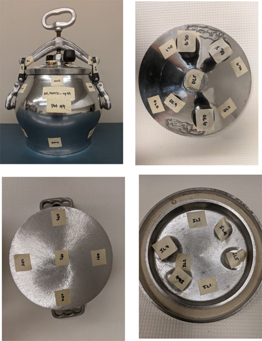
Investigating aluminum cookpots as a source of lead exposure

alexandrossfakianakis shared this article with you from Inoreader

41370_2022_431_Fig1_HTML.png

Journal of Exposure Science & Environmental Epidemiology, Published online: 02 May 2022; doi:10.1038/s41370-022-00431-y

Investigating aluminum cookpots as a source of lead exposure in Afghan refugee children resettled in the United States
View on the web

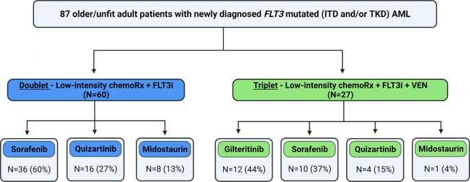
Hypomethylating agent and venetoclax with FLT3 inhibitor “triplet” therapy in older/unfit patients with FLT3 mutated AML

alexandrossfakianakis shared this article with you from Inoreader

41408_2022_670_Fig1_HTML.png

Blood Cancer Journal, Published online: 02 May 2022; doi:10.1038/s41408-022-00670-0

Hypomethylating agent and venetoclax with FLT3 inhibitor "triplet" therapy in older/unfit patients with FLT3 mutated AML
View on the web

Extravasation and outcomes in computed tomography and angiography in patients with pelvic fractures requiring transcatheter arterial embolization: A single-center observational study

alexandrossfakianakis shared this article with you from Inoreader
imageBACKGROUND Extravasation on contrast-enhanced computed tomography (CECT) is a helpful indicator of the need for transcatheter arterial embolization (TAE) for pelvic fractures. However, previous reports were inconsistent on cases in which angiography is necessary, even though there is no extravasation on computed tomography. This study aimed to describe and analyze the contradictory findings in cases where extravasation is observed on angiography but not on CECT, to contribute to improved management of patients with pelvic fractures. METHODS This was a retrospective single-center study. Patients with pelvic fractures who underwent CECT and TAE between 2014 and 2020 were included. We classified the patients into three groups: CECT and angiography with extravasation (CT+Angio+), CECT with no extravasation and angiography with extravasation (CT−Angio+), and CECT with extravasation and angiography without extravasation (CT+Angio−). RESULTS A total of 113 patients were included in the study: the CT+Angio+ group had 54 patients, CT−Angio+ group, 47; and CT+Angio− group, 12. The CT−Angio+ group had a significantly longer time from arrival to CECT than the CT+Angio+ group (27 minutes vs. 23 minutes, p
View on the web

Direct to OR resuscitation of abdominal trauma: An NTDB propensity matched outcomes study

alexandrossfakianakis shared this article with you from Inoreader
imageBACKGROUND Direct to operating room resuscitation (DOR) is used by some trauma centers for severely injured trauma patients as an approach to minimize time to hemorrhage control. It is unknown whether this strategy results in favorable outcomes. We hypothesized that utilization of an emergency department operating room (EDOR) for resuscitation of patients with abdominal trauma at an urban Level I trauma center would be associated with decreased time to laparotomy and improved outcomes. METHODS We included patients 15 years or older with abdominal trauma who underwent emergent laparotomy within 120 minutes of arrival both at our institution and within a National Trauma Data Bank sample between 2007 to 2019 and 2013 to 2016, respectively. Our institutional sample was matched 1:1 to an American College of Surgeons National Trauma Databank sample using propensity score matching based on age, sex, mechanism of injury, and abdominal Abbreviated Injury Scale score. The primary outcome was time to laparotomy incision. Secondary outcomes included blood transfusion requirement, intensive care unit (ICU) length of stay (LOS), ventilator days, hospital LOS, and in-hospital mortality. RESULTS Two hundred forty patients were included (120 institutional, 120 national). Both samples were well balanced, and 83.3% sustained penetrating trauma. There were 84.2% young adults between the ages of 15 and 47, 91.7% were male, 47.5% Black/African American, with a median Injury Severity Score of 14 (interquartile range [IQR], 8–29), Glasgow Coma Scale score of 15 (IQR, 13–15), 71.7% had an systolic blood pressure of >90 mm Hg, and had a shock index of 0.9 (IQR, 0.7–1.1) which did not differ between groups (p > 0.05). Treatment in the EDOR was associated with decreased time to incision (25.5 minutes vs. 40 minutes; p ≤ 0.001), ICU LOS (1 vs. 3.1 days; p
View on the web

Artificial Enamel – Stronger and Durable finds other uses beyond Medicine

alexandrossfakianakis shared this article with you from Inoreader

Artificial Enamel Developed in Lab stronger than Natural Enamel

Researchers have successfully designed artificial Enamel in their lab, which might not be used in repairing or replacing lost Enamel on natural teeth but can be used in other areas beyond medicine. Artificial Enamel can be used in creating Body armor, stronger body for Vehicles, hardening surfaces of many appliances and even in building materials which can help in protecting against Earthquakes as wel l. The Artificial or Lab grown Enamel has been found to out perform the natural enamel in six different areas such as Elasticity, absorbing vibrations, strength etc. Enamel is a Complex structure and to replicate it and mimic all its properties is a big step in the right direction. Natural Enamel is the hardest tissue in the human body which is strong enough to not crack on applying hard forces for lifetime and also elastic enough to bear all types of forces. Natural Enamel has many nested modes of organization like wool fibers spun into yarn and then knitted in to a cable knit sweater, Calcium, phosphorus and oxygen atoms must come together in a complex repeating pattern to form crystalline wires. To achieve artificial enamel structure, researchers used extreme temperature to arrange the wires in an orderly formation to achieve the required arrangement. Earlier scientists unsuccessfully used peptides - short chains of amino acids which help in building proteins to mimic enamel. With the new attempt Researchers used malleable metal based coating to encase the wires to give extra strength
Artificial Enamel Developed in Lab stronger than Natural Enamel

According to Nicholas Kotov, a chemical engineer at the University of Michigan, Ann Arbor, said - What makes the artificial Enamel more like Natural Enamel is the coating on the crystalline wires which makes the Artificial Enamel more resilient and less likely to break, the soft material covering the wires can absorb high amount of pressures and shock. The magnesium rich coating of the Natural Enamel has been replaced with Zirconium Oxide giving it more strength and also non toxic. The use of these materials helped in achieving a material which was hard and could be cut into desired shapes using a diamond based saw. The team of Researchers have tested the hardness and elasticity of the new Artificial Enamel material by making a small notch on the surface and applied pressure on it until it gave in and formed a fracture. The toughness of the material was determined by seeing the pressure applied and the length of the crack, it also helped in determining the strain resistance of the enamel. The researchers even tried Artificial Enamel against Natural Enamel where the results where in favor of lab grown enamel outperforming the Natural one. The cells (Ameloblasts) which form Enamel die as soon as the tooth emerges from the gums hence Enamel cannot repair itself. Most dental visits are due to Caries in the Enamel and n neglecting, it might lead to tooth loss as well. To replace damaged Enamel, Dentists use artificial filling materials such as GIC, Composite, Dental Amalgam, etc which do not have the same properties such as Natural Enamel to withstand the Oral forces for decades. Use of this Artificial Enamel in repairing natural Enamel might still take some time as bonding Artificial Enamel to Natural Enamel needs to first heated to 300 degrees C first, Frozen and then cut into the required shape which is not so easy in a Clinical practice. References: 
View on the web

Δημοφιλείς αναρτήσεις