Τετάρτη 14 Απριλίου 2021

Cancers, Vol. 13, Pages 1891: Tumor Solid Stress: Assessment with MR Elastography under Compression of Patient-Derived Hepatocellular Carcinomas and Cholangiocarcinomas Xenografted in Mice

xloma.fota13 shared this article with you from Inoreader
Via Cancers

cancers-13-01891-ag-550.jpg

Cancers, Vol. 13, Pages 1891: Tumor Solid Stress: Assessment with MR Elastography under Compression of Patient-Derived Hepatocellular Carcinomas and Cholangiocarcinomas Xenografted in Mice

Cancers doi: 10.3390/cancers13081891

Authors: Gwenaël Pagé Marion Tardieu Jean-Luc Gennisson Laurent Besret Philippe Garteiser Bernard E. Van Beers

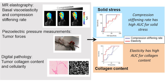
Malignant tumors have abnormal biomechanical characteristics, including high viscoelasticity, solid stress, and interstitial fluid pressure. Magnetic resonance (MR) elastography is increasingly used to non-invasively assess tissue viscoelasticity. However, solid stress and interstitial fluid pressure measurements are performed with invasive methods. We studied the feasibility and potential role of MR elastography at basal state and under controlled compression in assessing altered biomechanical features of malignant liver tumors. MR elastography was performed in mice with patient-derived, subcutaneously xenografted hepatocellular carcinomas or cholangiocarcinomas to measure the basal viscoelasticity and the compression stiffening rate, which corresponds to the slope of elasticity versus applied compression. MR elastography measurements were correlated with invasive pressure measurements and digital histological readings. Significant differences in MR elastography parameters, pres sure, and histological measurements were observed between tumor models. In multivariate analysis, collagen content and interstitial fluid pressure were determinants of basal viscoelasticity, whereas solid stress, in addition to collagen content, cellularity, and tumor type, was an independent determinant of compression stiffening rate. Compression stiffening rate had high AUC (0.87 ± 0.08) for determining elevated solid stress, whereas basal elasticity had high AUC for tumor collagen content (AUC: 0.86 ± 0.08). Our results suggest that MR elastography compression stiffening rate, in contrast to basal viscoelasticity, is a potential marker of solid stress in malignant liver tumors.

View on the web

Τρίτη 13 Απριλίου 2021

Rhinosurgery during and after the COVID-19 Pandemic: International Consensus Conference Statement on Preliminary Perioperative Safety Measures

xloma.fota13 shared this article with you from Inoreader
imageBackground: The coronavirus disease of 2019 (COVID-19) pandemic has widely affected rhinosurgery, given the high risk of contagion and the elective nature of the aesthetic procedure, generating many questions on how to ensure safety. The Science and Research Committee of the Rhinoplasty Society of Europe aimed at preparing consensus recommendations on safe rhinosurgery in general during the COVID-19 pandemic by appointing an international panel of experts also including delegates of The Rhinoplasty Society. Methods: A Zoom meeting was performed with a panel of 14 international leading experts in rhinosurgery. During 3.5 hours, four categories of questions on preoperative safety measures in private practice and outpatient clinics, patient assessment before and during surgery, and legal issues were presented by four chairs and discussed by the expert group. Afterward, the panelists were requested to express an online, electronic vote on each category and question. The panel's recommendations were based on current evidence and expert opinions. The resulting report was circled in an iterative open e-mail process until consensus was obtained. Results: Consensus was obtained in several important points on how to safely restart performing rhinosurgery in general. Preliminary recommendations with different levels of agreement were prepared and condensed in a bundle of safety measures. Conclusion: The implementation of the panel's recommendations may improve safety of rhinoplasty by avoiding operating on nondetected COVID-19 patients and minimizing severe acute respiratory syndrome coronavirus 2 virus spread in outpatient clinics and operating rooms.
View on the web

The Utility of the Subalar Graft in Nostril Symmetry in Rhinoplasty

xloma.fota13 shared this article with you from Inoreader
imageBackground: A deviated nose can be attributable to multiple anatomical factors, including asymmetric maxilla. A subalar graft helps to correct maxillary hypoplasia and may be a useful tool for correcting a deviated nose. The authors' objective is to show the effects of the subalar graft in improving nostril symmetry and to propose an algorithm for using this graft in open and endonasal rhinoplasty. Methods: A retrospective case series was performed on patients who had undergone rhinoplasty performed by the senior author (R.W.) from September of 2008 to July of 2015. Patients with at least 3 months of follow-up and adequate photographs were included. The mean follow-up period was 11.0 months (range, 3 to 72 months). A total of 68 patients were included. Preoperative and postoperative photographs were analyzed to measure changes in nasal axis deviation, alar facial angle on base view, alar facial angle on frontal view, and nostril show bilaterally. Results: Of the 68 patients, statistically significant improvement of nasal axis deviation of 4.32 degrees toward the midline was observed. Alar facial angle on base view was improved 1.01 degrees toward the horizontal. Nostril symmetry also improved based on the ratio between the shorter side and the longer side. The mean change in nostril show ratio was 0.19 toward a 1:1 ratio. Conclusions: Previous studies have shown that the subalar grafting technique is an important adjunctive technique in rhinoplasty for patients with midfacial asymmetries. This case series demonstrates that this technique can provide sustained results in the correction of the nasal foundation. CLINICAL QUESTION/LEVEL OF EVIDENCE: Therapeutic, IV.
View on the web

The Association of Zygomaticomaxillary Complex Fractures with Naso-Orbitoethmoid Fractures in Pediatric Populations

xloma.fota13 shared this article with you from Inoreader
imageBackground: Naso-orbitoethmoid fractures associated with ipsilateral zygomaticomaxillary complex fractures are more challenging injuries than zygomaticomaxillary complex fractures alone. However, there is a paucity of information on this complex fracture pattern in the pediatric population. This study investigated the cause, treatment, and outcomes of combined zygomaticomaxillary complex and naso-orbitoethmoid fractures versus isolated zygomaticomaxillary complex fractures in pediatric patients. Methods: This was a 25-year retrospective cohort study of pediatric patients who presented to a single institution with zygomaticomaxillary complex fractures. Baseline patient demographics and clinical information, and concomitant injuries, treatment/operative management, and postoperative complications/deformities were recorded and compared between patients with combined zygomaticomaxillary complex and naso-orbitoethmoid fractures and patients with isolated zygomaticomaxillary complex fractures. Results: Forty-nine patients were identified to have had zygomaticomaxillary complex fractures in the authors' 25-year study period, of whom 46 had adequate clinical documentation and follow-up. Seventeen patients had combined zygomaticomaxillary complex–naso-orbitoethmoid fractures, of whom six had panfacial fractures. Both patient groups (zygomaticomaxillary complex only and combined zygomaticomaxillary complex–naso-orbitoethmoid fractures) were similar in terms of demographics. However, a significantly greater proportion of combined fracture patients experienced postoperative complications compared to isolated zygomaticomaxillary complex fracture patients, even after excluding those with panfacial fractures (87.5 percent versus 35.3 percent; p
View on the web

The Intra.Ox Near-Infrared Spectrometer Measures Variations in Flap Oxygenation That Correlate to Flap Necrosis in a Preclinical Rodent Model

xloma.fota13 shared this article with you from Inoreader
imageBackground: Mastectomy flap necrosis affects 7 to 40 percent of patients undergoing immediate breast reconstruction, with many cases resulting in infection and/or explantation. The Intra.Ox near-infrared spectrometer is one of several novel devices that assesses tissue perfusion by measuring the interactions of light with oxygenated and deoxygenated hemoglobin. This handheld device facilitates serial flap perfusion assessment and may objectively identify at-risk tissues and guide evidence-based treatment algorithms. In this preliminary study, we hypothesized that the Intra.Ox spectrometer detects differences in tissue oxygenation that correlate to tissue necrosis. Methods: Dorsal, random-pattern flaps measuring 10 × 3 cm were raised in eight male Sprague-Dawley rats. Intraoperative tissue oxygen saturation was measured using Intra.Ox in 10 standardized locations. On postoperative day 7, the skin flaps were evaluated for full-thickness necrosis. Data were analyzed using the chi-square test and one-way analysis of variance. A receiver operating characteristic curve assessed the accuracy of intraoperative tissue oxygenation in predicting the risk of flap necrosis. Results: Tissue oxygen saturation exhibited a strong negative correlation to distance from the flap pedicle (r = −0.798). Oxygen saturation in tissue that developed necrosis averaged 32 percent, compared to 59 percent in tissues that did not (p
View on the web

Impacts of Rehabilitation Gait Training on Functional Outcomes after Tibial Nerve Transfer for Patients with Peroneal Nerve Injury: A Nonrandomized Controlled Trial

xloma.fota13 shared this article with you from Inoreader
imageBackground: Although there was initial success using tibial nerve transfer to restore ankle dorsiflexion following peroneal nerve injury, results from later series were less promising. A potential reason is coactivation of the much stronger antagonistic muscles during gait. The purpose of this study was to test the hypothesis that gait training would improve functional performance following tibial nerve transfer. Methods: Using a prospective, nonrandomized, controlled study design, patients were divided into two groups: surgery only or surgery plus gait training. Of the 20 patients who showed reinnervation in the tibialis anterior muscle, 10 were assigned to the gait training group, and an equal number were in the control group. Those in the treatment group began training once reinnervation in the tibialis anterior muscle was detected, whereas those in the control group continued to use their ankle-foot orthosis full time. Differences in ankle dorsiflexion was measured using the Medical Research Council scale, and quantitative force measurement and functional disability was measured using the Stanmore Scale. Results: Patients in the gait training group attained significantly better functional recovery as measured by the Stanmore Scale (79.5 ± 14.3) (mean ± SD) versus (37.2 ± 3.5) in the control group (p = 0.02). Medical Research Council grades were 3.8 ± 0.6 in the training group versus 2.5 ± 1.2 in the surgery only group (p
View on the web

Cone-Beam Computed Tomography: A User-Friendly, Practical Roadmap to the Planning and Execution of Every Rhinoplasty—A 5-Year Review

xloma.fota13 shared this article with you from Inoreader
imageBackground: Cone-beam computed tomography has recently rapidly developed worldwide as a versatile and convenient alternative to traditional computed tomography for imaging of the maxillofacial region. However, most surgeons performing rhinoplasty are surprisingly unfamiliar with it, in both the plastic surgery and ear, nose, and throat communities. Methods: The broad clinical experience of a single center over the past 5 years is reviewed. The many applications of cone-beam computed tomography to primary and secondary rhinoplasty are analyzed regarding septum, turbinates, nasal bones, skin thickness, and other issues. The importance of a paradigm shift from a two-dimensional to a three-dimensional approach in image reconstruction is demonstrated, together with the value of surface contour enhancement. Results: Cone-beam computed tomography has a multitude of practical applications highly relevant to rhinoplasty. The surface image will strengthen aesthetic analysis, and the detailed preview of bony and functional anatomy will facilitate surgical planning. Cone-beam computed tomography serves as a roadmap to plan and execute rhinoplasties more predictably and efficiently. The availability of spatial views and accurate detail, together with the possibility of easy, accurate measuring, offers a plenitude of potential applications. Conclusions: Cone-beam computed tomography is a user-friendly, quick technique with abundant advantages in planning any rhinoplasty. It causes the patient no inconvenience and has very few, if any, drawbacks, with these being limited to radiation exposure and limited cost.
View on the web

Δημοφιλείς αναρτήσεις