Πέμπτη 4 Φεβρουαρίου 2021

An Enlarging Parotid Mass in a 9-Year-Old Boy

xlomafota13 shared this article with you from Inoreader

ocg200023f1_1612213508.21079.png?Expires

A 9-year-old boy presents with a left-sided neck mass that has been enlarging, mild intermittent pain, episodes of upper respiratory infection, and previous adenotonsillectomy and bilateral ear tubes placement. What is your diagnosis?
View on the web

Povidone Iodine to Reduce Viral Load in Patients With COVID-19

xlomafota13 shared this article with you from Inoreader

old200020t1_1612213510.49203.png?Expires

This randomized clinical trial investigates the efficacy of na sopharyngeal povidone iodine solutions in reducing the viral load of patients with COVID-19.
View on the web

Diffuse Type 2 Dominant Rhinosinusitis and Corticosteroid Irrigation After Surgical Neosinus Cavity Formation

xlomafota13 shared this article with you from Inoreader

ooi200092f1_1612213519.55448.png?Expires

This cohort study examines long-term treatment outcomes in pat ients with eosinophilic rhinosinusitis who have received corticosteroid irrigations after surgical creation of a neosinus cavity.
View on the web

New Age Mentoring and Disruptive Innovation

xlomafota13 shared this article with you from Inoreader

osc200003f1_1612213523.60132.png?Expires

This study examines how mentoring has evolved over time, what new challenges have recently emerged, and how to implement effective structural solutions in the field of otolaryngology–head and neck surgery.
View on the web

Risk and Clinical Factors of Late Lower Cranial Neuropathy in Oropharyngeal Squamous Cell Carcinoma

xlomafota13 shared this article with you from Inoreader

ooi200091f1_1612213513.57013.png?Expires

This cohort study assesses the cumulative incidence of and cli nical factors associated with late lower cranial neuropathy among patients with long-term survival of oropharyngeal squamous cell carcinoma.
View on the web

Prognostic Significance of Systemic Inflammatory Response in Cases of Temporal Bone Squamous Cell Carcinoma

xlomafota13 shared this article with you from Inoreader

Objective/Hypothesis

Squamous cell carcinoma (SCC) of the temporal bone is an extremely rare condition. This rarity has led to a delay in the establishment of a standard treatment protocol and adequate staging system. Identification of prognostic markers of this disease from a variety of fields is desirable in the establishment of treatment guidelines for temporal bone SCC. The aim of this study is to assess the prognostic role of inflammation‐based prognostic scores in cases of temporal bone SCC.

Study Design

Case reries with chart review.

Methods

A total of 71 cases of primary malignancy eligible for curative treatment at a single tertiary medical institute were retrospectively analyzed. Univariate and multivariate regression analyzes were used to investigate the association between the inflammation‐based scores and 5‐year overall survival.

Results

Univariate Cox regression analyzes showed that a high neutrophil‐to‐lymphocyte ratio, high platelet‐to‐lymphocyte ratio, low lymphocyte‐to‐monocyte ratio, a Glasgow prognostic score of 2, and the systemic inflammation score of 2 were significantly associated with a poor prognosis, as well as a classification of T4 stage, presence of cervical lymph node metastasis, high white blood cell counts, and high C‐reactive protein levels. The multivariate analysis showed that a clinical stage of T4 and a systemic inflammation score of 2 were independent prognostic markers.

Conclusions

Inflammation‐based prognostic markers are associated with the survival of patients with temporal bone SCC, as well as other head and neck SCCs.

Level of Evidence

4 Laryngoscope, 2021

View on the web

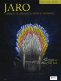
Residual Hair Cell Responses in Electric-Acoustic Stimulation Cochlear Implant Users with Complete Loss of Acoustic Hearing After Implantation

xlomafota13 shared this article with you from Inoreader

10162.jpg

Abstract

Changes in cochlear implant (CI) design and surgical techniques have enabled the preservation of residual acoustic hearing in the implanted ear. While most Nucleus Hybrid L24 CI users retain significant acoustic hearing years after surgery, 6–17 % experience a complete loss of acoustic hearing (Roland et al. Laryngoscope. 126(1):175-81. (2016), Laryngoscope. 128(8):1939-1945 (2018); Scheperle et al. Hear Res. 350:45-57 (2017)). Electrocochleography (ECoG) enables non-invasive monitoring of peripheral auditory function and may provide insight into the pathophysiology of hearing loss. The ECoG response is evoked using an acoustic stimulus and includes contributions from the hair cells (cochlear microphonic—CM) as well as the auditory nerve (auditory nerve neurophonic—ANN). Seven Hybrid L24 CI users with complete loss of residual hearing months after surgery underwent ECoG measures before and after loss of hearing. While significant reductions in CMs were evident after hearing loss, all participants had measurable CMs despite having no measurable acoustic hearing. None retained measurable ANNs. Given histological data suggesting stable hair cell and neural counts after hearing loss (e.g., Quesnel et al. Hear Res. 333:225-234. (2016)), the loss of ECoG and audiometric hearing may reflect reduced synaptic input. This is consistent with the theory that residual CM responses coupled with little to no ANN responses reflect a "disconnect" between hair cells and auditory nerve fibers (Fontenot et al. Ear Hear. 40(3):577-591. 2019). This "disconnection" may prevent proper encoding of auditory stimulation at higher auditory pathways, leading to a lack of audiometric responses, even in the presence of viable cochlear hair cells.

View on the web

Salivary Duct Carcinoma With Rhabdoid Features—No or Aberrant Expression of E-cadherin and Genetic Changes in CDH1: Immunohistochemical and Genetic Analyses of 17 Cases

xloma.fota13 shared this article with you from Inoreader

SocialThumb.00000478.DC.jpeg

Salivary duct carcinoma is a relatively uncommon malignancy of the salivary glands; however, it frequently occurs as a carcinomatous component of carcinoma ex pleomorphic adenoma. We previously reported salivary duct carcinoma with rhabdoid features (SDCRF) as an extremely rare subtype of salivary duct carcinoma, and that it occurred as a salivary counterpart of pleomorphic lobular carcinoma of the breast (PLCB). We collected new cases of SDCRF for this study, in which we examined a total of 17 cases immunohistochemically and genetically. As it is known that PLCB exhibits loss of or aberrant E-cadherin expression and carries nonsense/missense mutations in or deletion of the CDH1 gene, we examined the CDH1 gene status of our SDCRF cases. All of the examined SDCRF cases involved the diffuse proliferation of large ovoid cells with eosinophilic cytoplasm and eccentric nuclei, which displayed reduced cell-cell adhesion. Most cases were positive for pan-cytokeratin, androgen receptor, gross cystic disease fluid protein-15, SWI/SNF-related matrix-associated actin-dependent regulator of chromatin subfamily B member 1, and WI/SNF-related matrix-associated actin-dependent regulator of chromatin subfamily A member 4, whereas they were negative for vimentin. No and decreased/cytoplasmic E-cadherin expression was ob served in 11 and 4 of 17 cases, respectively, whereas no and decreased/cytoplasmic β-catenin expression were observed in 10 and 5 of 17 cases, respectively. Among the 11 cases that could be genetically analyzed, a nonsense mutation (1 case), missense mutations (6 cases), and insertions (1 case) were detected in the CDH1 gene. In conclusion, we propose that SDCRF is the salivary counterpart of PLCB due to its morphology and immunophenotype, and the genetic status of CDH1. This study was approved by the Institutional Review Board of Shizuoka General Center (SGHIRB#2019007) and Hamamatsu University School of Medicine (20-011). All subjects signed informed consent forms. Written informed consent for the publication of clinical details and/or clinical images was obtained from the patients. A copy of the consent form is available for review by the Editor of this journal. K.K.: designed and drafted the manuscript. K.K. and M.S.: made the histopathologic diagnoses. M.M., K.Y., S.B., T.O., and H.I.: found the cases and collected the clinical data. H. Yamamoto, T.I., and C.T.: performed the immunohistochemistry. H. Yamada and K.I.: performed the genetic analysis. H.S. and M.S.: supervised this manuscript. Conflicts of Interest and Source of Funding: Supported in part by a Grant-in-Aid for the Medical Research Support Project of Shizuoka Prefectural Hospital Organization in 2019 (to K.K.), as well as by AMED (20ck0106554) (to H.S.) and SRF (to H.S.). The authors have disclosed that they have no significant relationships with, or financial interest in, any commercial companies pertaining to this article. Correspondence: Kimihide Kusafuka, DDS, PhD, Department of Pathology, Shizuoka General Hospital, 4-27-1 Kita-Ando, Aoi-ku, Shizuoka City 420-8527, Shizuoka Prefecture, Japan (e-mail: k-kusafuka@i.shizuoka-pho.jp). Copyright © 2021 Wolters Kluwer Health, Inc. All rights reserved.
View on the web

A Distinctive Adnexal (Usually Paratubal) Neoplasm Often Associated With Peutz-Jeghers Syndrome and Characterized by STK11 Alterations (STK11 Adnexal Tumor): A Report of 22 Cases

xloma.fota13 shared this article with you from Inoreader

SocialThumb.00000478.DC.jpeg

We describe 22 examples of a novel, usually paratubal, adnexal tumor associated with Peutz-Jeghers syndrome in nearly 50% of cases that harbored STK11 alterations in all tested (n=21). The patients ranged from 17 to 66 years (median=39 y) and the tumors from 4.5 to 25.5 cm (median=11 cm). Most (n=18) were paratubal, wi th metastases noted in 11/22 (50%) and recurrences in 12/15 (80%). Morphologically, they were characterized by interanastomosing cords and trabeculae of predominantly epithelioid cells, set in a variably prominent myxoid to focally edematous stroma, that often merged to form tubular, cystic, cribriform, and microacinar formations, reminiscent of salivary gland-type tumors. The tumor cells were uniformly atypical, often with prominent nucleoli and a variable mitotic index (median=9/10 HPFs). The tumors were usually positive to a variable extent for epithelial (CAM5.2, AE1/AE3, cytokeratin 7), sex cord (calretinin, inhibin, WT1), and mesothelial (calretinin, D2-40) markers, as well as hormone receptors. PAX8, SF1, and GATA-3 were rarely positive, while claudin-4, FOXL2, and TTF-1 were consistently negative. All sequenced tumors (n=21) harbored alterations in STK11, often with a loss of heterozygosity event. There were no other recurrently mutated genes. Recurrent copy number alterat ions included loss of 1p and 11q, and gain of 1q, 15q, and 15p. Despite an extensive morphologic, immunohistochemical, and molecular evaluation, we are unable to determine with certainty the histogenesis of this unique tumor. Wolffian, sex cord stromal, epithelial, and mesothelial origins were considered. We propose the term STK11 adnexal tumor to describe this novel entity and emphasize the importance of genetic counseling in these patients as a significant number of neoplasms occur in association with Peutz-Jeghers syndrome. Conflicts of Interest and Source of Funding: B.W. was funded in part by Breast Cancer Research Foundation, Cycle for Survival and Stand Up To Cancer grants. Research reported in this publication was supported in part by a Cancer Center Support Grant of the NIH/NCI (Grant No. P30CA008748; MSKCC). The authors have disclosed that they have no significant relationships with, or financial interest in, any commercial companies pertaining to this article. Correspondence: Jennifer A. Bennett, MD, Department of Pathology, University of Chicago Medicine, 5841 South Maryland Avenue, MC6101, Chicago, IL 60637 (e-mail: jabennett@bsd.uchicago.edu). Copyright © 2021 Wolters Kluwer Health, Inc. All rights reserved.
View on the web

OGT-rearranged Acral Mesenchymal Neoplasms: An Emerging Entity With an Expanding Pathologic and Molecular Spectrum

xloma.fota13 shared this article with you from Inoreader

SocialThumb.00000478.DC.jpeg

No abstract available
View on the web

Objective Visual Analog Scale for Biopsy Diagnosis of Helicobacter pylori Infection in Clinical Practice

xloma.fota13 shared this article with you from Inoreader

SocialThumb.00000478.DC.jpeg

Historic and current pathology society guidelines recommend using visual gestalt to identify substantial inflammatory cell infiltrate in Helicobacter pylori gastritis, but these scales were subjectively designed. This study aims to objectively investigate the density of inflammation that justifies additional workup for H. py lori infection. We retrospectively identified 2 patient cohorts who had undergone endoscopy with gastric biopsies; 1 with H. pylori infection (n=66), confirmed with a positive stool antigen test and/or Campylobacter-like organism test, and 1 without infection (n=81). Antral and body biopsies were selected from each case, if available, and stained with MUM-1 to highlight mucosal plasma cells. Digital analysis was performed to calculate the number of plasma cells/mm2, termed the "inflammatory score" (IS). Patients with H. pylori infection had an average of 1289 plasma cells/mm2 in the antrum and 835 plasma cells/mm2 in the body, compared with 346 plasma cells/mm2 in the antrum and 178 plasma cells/mm2 in the body in patients without infection. IS cut-off values for a positive infection were 714 plasma cells/mm2 in the antrum and 316 plasma cells/mm2 in the body, with high sensitivities and specificities in both the antrum (92%, 92%) and body (85%, 84%), respectively. A visual anal og scale was created to provide a histologic correlate of the observed IS ranges and cut-offs. This practical and objective scale is associated with a high sensitivity and specificity for diagnosing H. pylori infection and justifies moving away from upfront universal H. pylori testing in routine clinical practice. Conflicts of Interest and Source of Funding: The authors have disclosed that they have no significant relationships with, or financial interest in, any commercial companies pertaining to this article. Correspondence: Katherine E. Boylan, MD, Department of Anatomic Pathology, The University of Utah, 2000 Circle of Hope, Rm 3100, Salt Lake City, UT 84112 (e-mail: katherine.boylan@hsc.utah.edu). Copyright © 2021 Wolters Kluwer Health, Inc. All rights reserved.
View on the web

Δημοφιλείς αναρτήσεις