Κυριακή 29 Αυγούστου 2021

Nivolumab exposure in a hemodialysis patient with metastatic melanoma

xlomafota13 shared this article with you from Inoreader

SocialThumb.00008390.DC.jpeg

The effect of intermittent hemodialysis (IHD) on nivolumab serum concentrations in patients with severe renal impairment is largely unknown. Here, we present a 79-year-old patient with metastatic melanoma and end-stage renal disease on IHD three times a week, treated with 480 mg nivolumab every 4 weeks. A serum trough concentration of nivolumab was determined before the start of the third cycle, and two samples were taken immediately before and after a hemodialysis session during this cycle. All nivolumab serum concentrations were within a similar range as those previously measured among patients without renal insufficiency, after a comparable duration of nivolumab treatment. Therefore, we conclude that IHD does not influence nivolumab exposure. Furthermore, nivolumab treatment was continued without complications and appears to be well tolerated for patients on IHD. Received 22 March 2021 Accepted 24 July 2021 Correspondence to Robin van Geel, PharmD, PhD, Department of Clinical Pharmacy & Toxicology, Maastricht University Medical Center, P. Debyelaan 25, 6229 HX the Netherlands, Tel: +0031 043 3871881; e-mail: robin.van.geel@mumc.nl Copyright © 2021 Wolters Kluwer Health, Inc. All rights reserved.
View on the web

Expression of the cancer stem cell marker OCT4 is associated with worse prognosis and survival in cutaneous melanoma

xlomafota13 shared this article with you from Inoreader

SocialThumb.00008390.DC.jpeg

Cutaneous melanoma has an aggressive clinical presentation, showing rapid rate of growth and metastatic dissemination due to the permanence of cancer stem cells. The present study was to evaluate the expression of the self-renewal regulatory factor and the clinical significance of the transcription factor OCT4 in me lanoma. Melanoma tissues were stained by immunohistochemistry and the correlation between the expression of this marker was determined through clinical–pathological variables and survival outcomes. Positive expression of nuclear and cytoplasmic OCT4 was observed in 49% and 41.2% of cases, respectively. The positive expression of nuclear OCT4 in melanoma was significantly associated with prognostic factors, such as Breslow depth, Clark's level, ulceration and metastasis. Survival of patients was 56% compared to positive nuclear OCT4 expression and 94.2% when compared to the low expression of the gene. Nuclear OCT4 positive genotype indicated aggressive tumor behavior with a worse clinical outcome, which indicates OCT4 as a useful biomarker in the prognosis of melanoma. Received 10 November 2020 Accepted 1 July 2021 Correspondence to Kleber Santiago Freitas e Silva, PhD, Department of Medicine, Anápolis University Center, UniEvangélica, Avenida Universitária, Km 3,5 - Cidade Universitária, CEP 75083-515 Anápolis, Goiás, Brazil, Tel: +55 (62) 99924 5983; e-mail: smallbinho@hotmail.com Copyright © 2021 Wolters Kluwer Health, Inc. All rights reserved.
View on the web

COVID-19 vaccination mimicking lymph-node progression in a patient with melanoma: a case report

xlomafota13 shared this article with you from Inoreader

SocialThumb.00008390.DC.jpeg

COVID-19 vaccination has been rapidly implemented among patients with cancer. We present the case of a patient with high-risk resected cutaneous melanoma, who was a candidate for adjuvant treatment, with postsurgery 18-fluorodeoxyglucose (FDG) PET/computed tomography (CT) scan showing positive axillary lymph nodes after COV ID-19 vaccination. This report presents a 50-year-old man with a history of stage IIA cutaneous melanoma. During follow-up, the patient experienced subcutaneous and lymph-node disease progression, documented with 18FDG PET/CT scan. The patient underwent laparoscopic left para-aortic lymphadenectomy and excision of subcutaneous lesion. Histologic examination showed presence of melanoma metastases in 2 lymph nodes out of total 17 excised and neoplastic emboli to the subcutaneous tissue. In view of starting adjuvant nivolumab, the patient underwent CT scan restaging, with evidence of suspect centimetric periaortic and paracaval lymph nodes, which were deemed worthy of 18FDG PET investigation. The 18FDG PET/CT was negative for abdominal hypercaptation, but showed left axillary pathologic lymph nodes. The medical history of the patient revealed that he had received intramuscular Moderna COVID-19 mRNA vaccine in the left deltoid, one week before 18FDG PET examination. Since the patient' s clinical examination was negative and suspecting postvaccination false-positive adenopathy, bilateral axillary ultrasound was performed, excluding the presence of pathologic lymph nodes. The patient has started adjuvant treatment with nivolumab, which is currently ongoing. This case demonstrates unexpected findings in response to COVID-19 vaccination in a patient with melanoma. In this specific case, the detection of 18FDG PET hypercaptation could significantly change the patient's management. With growing evidence about the pattern and occurrence of adenopathies after mRNA COVID-19 vaccination, recommendations for scheduling and interpretation of 18FDG PET/CT scans among cancer patients will be implemented, in order to reduce equivocal findings and improve outcomes. Received 12 May 2021 Accepted 3 July 2021 Correspondence to Alice Indini, MD, Medical Oncology Unit, Department of Internal Medicine, Fondazione IRCCS Ca' Granda, Ospedale Maggiore Policlinico, via Francesco Sforza 35, 20122 Milano, Italy, Tel: +39 0255032660; fax: +39 0255032659; e-mail: alice.indini@gmail.com Copyright © 2021 Wolters Kluwer Health, Inc. All rights reserved.
View on the web

Aseptic cystitis induced by nivolumab and ipilimumab combination for metastatic melanoma

xlomafota13 shared this article with you from Inoreader

SocialThumb.00008390.DC.jpeg

Immune checkpoint inhibitors (ICIs) have revolutionized the treatment of advanced melanoma. Combination of ICI with ipilimumab cytotoxic T-lymphocyte antigen-4 and nivolumab [anti-programmed cell death-1 (PD-1)] improves tumoral response compared to anti-PD1 monotherapy in melanoma patients, but is associated with more seve re and multiple immune-related adverse events. We report the first case of aseptic cystitis induced by ipilimumab and nivolumab combination in a 61-year-old melanoma patient. She described after two infusions, diarrhea, pollakiuria, intense bladder pain, urinary urgency, and nocturia. Repeated negative urine culture tests led to perform cystoscopy. Mucosal bladder biopsies showed lymphocytic T-cells infiltration in intraepithelial and in subepithelial connective tissue, which were consistent with the diagnosis of immune-related aseptic cystitis. Aseptic cystitis is a rare and poorly known side-effect related to ICI. Only four other cases with anti-PD1 monotherapy were found in literature, only in Japanese patients. It simulates bacterial cystitis with negative urinary tests, and is often associated with atypical symptoms like diarrhea, which may delay the diagnosis. Oral steroids appear to be the most efficient therapeutic options. Received 4 May 2021 Accepted 28 June 2021 Correspondence to Sorilla Prey, MD, PhD, Service de Dermatologie, Groupe hospitalier Saint André, 1, rue Jean Burguet, F-33000 Bordeaux, France, Tel: +33 5 56 79 47 05; fax: +33 5 56 79 49 75; e-mail: sorilla.prey@chu-bordeaux.fr Copyright © 2021 Wolters Kluwer Health, Inc. All rights reserved.
View on the web

Metastatic acral melanoma treatment outcomes: a systematic review and meta-analysis

xlomafota13 shared this article with you from Inoreader

SocialThumb.00008390.DC.jpeg

Acral melanomas are a unique subset of melanomas occurring on the palms, soles, and nails. There is poor prognosis with surgery alone and no specific guidelines for the treatment of metastatic acral melanoma. This meta-analysis explored the systemic therapy outcomes for metastatic acral melanoma. Medline, Pubmed, EMBA SE, and the grey literature were searched from 2010 to August 2020 for studies specifying the treatment outcome of metastatic acral melanoma. Studies were assessed by two investigators. A random-effects meta-analysis was performed and pooled Kaplan–Meier curves for progression-free survival and overall survival were created. Critical appraisal was performed using the Newcastle-Ottawa Scale. Nineteen nonrandomized studies were included, comprising 646 patients with acral melanomas and 1609 patients with nonacral melanomas treated with systemic therapy including chemotherapy, KIT-targeted drugs, as well as anti-CTLA-4 and anti-PD-1 checkpoint inhibitor therapy. Thirteen studies included Kaplan–Meier curves for progression-free survival or overall survival and 11 studies reported treatment responses. Patients with acral melanomas had worse prognosis than nonacral cutaneous melanoma (acral overall survival: median 15 months, 95% CI, 13.7–16.3 months; nonacral cutaneous: median 24 months, 95% CI, 22.6–25.4 months, P 
View on the web

Clinical implications of alpha, beta, and gamma HPV infection in juvenile onset recurrent respiratory papillomatosis

xlomafota13 shared this article with you from Inoreader

Eur Arch Otorhinolaryngol. 2021 Aug 28. doi: 10.1007/s00405-021-07040-9. Online ahead of print.

ABSTRACT

PURPOSE: The aim of our study was to evaluate the prevalence of different HPV genera-alpha, beta and gamma-in Juvenile onset Recurrent Respiratory Papillomatosis (JoRRP) and examine the association of type and genus-specific viral features with the clinical outcome of disease.

METHODS: This retrospective observational study included consecutive patients with JoRRP who were treated in a referral centre between October 2000 and October 2020. All patients underwent cold excision and laser vaporisation of papillomatous lesions. Samples were analysed for the presence of 120 viral genotypes (22 alpha-HPV, 46 beta-HPV, 52 gamma-HPV) using a highly sensitive multiplex genotyping assay.

RESULTS: Twenty patients with JoRRP, aged 0.3-11 years, were included, with a median follow-up of 13.5 years. All samples were HPV DNA positive: 20 (100%) for alpha-HPV DNA; 7 (35%) for beta-HPV DNA; 0 for gamma-HPV DNA. Three groups were defined according to the number of infections: seven cases (35%) with HPV mono-infection; ten cases (50%) with HPV double-infection; three cases (15%) with ≥ 3 HPV infections. At diagnosis, patients with ≥ 3 HPV infections reported higher median Derkay's score than those with mono-infection (21 vs 14, P = 0.018). Number of HPV infections was also associated with clinical outcomes, with an average of 0.5 surgical procedures/year in patients with mono-infection, 1.2 for double-infection, 2.6 for ≥ 3 infections (P = 0.006).

CONCLUSION: Despite the small sample size, these preliminary data support an association between the number of different alpha and beta HPV co-infections and the clinical severity of the disease.

PMID:34453571 | DOI:10.1007/s00405-021-07040-9

View on the web

Adenoidectomy for middle ear disease in cleft palate children: a systematic review

xlomafota13 shared this article with you from Inoreader

Eur Arch Otorhinolaryngol. 2021 Aug 28. doi: 10.1007/s00405-021-07035-6. Online ahead of print.

ABSTRACT

PURPOSE: Cleft palate children have a higher incidence of otitis media with effusion, more frequent recurrent acute otitis media episodes, and worse conductive hearing losses than non-cleft children. Nevertheless, data on adenoidectomy for middle ear disease in this patient group are scarce, since many feared worsening of velopharyngeal insufficiency after the procedure. This review aims at collecting the available evidence on this subject, to frame possible further areas of research and interventions.

METHODS: A PRISMA-compliant systematic review was performed. Multiple databases were searched with criteria designed to include all studies focusing on the role of adenoidectomy in treating middle ear disease in cleft palate children. After duplicate removal, abstract and full-text selection, and quality assessment, we reviewed eligible articles for clinical indications and outcomes.

RESULTS: Among 321 unique citations, 3 studies published between 1964 and 1972 (2 case series and a retrospective cohort study) were deemed eligible, with 136 treated patients. The outcomes were positive in all three articles in terms of conductive hearing loss improvement, recurrent otitis media episodes reduction, and effusive otitis media resolution.

CONCLUSION: Despite promising results, research on adenoidectomy in treating middle ear disease in the cleft population has stopped in the mid-Seventies. No data are, therefore, available on the role of modern conservative adenoidectomy techniques (endoscopic and/or partial) in this context. Prospective studies are required to define the role of adenoidectomy in cleft children, most interestingly in specific subgroups such as patients requiring re-tympanostomy, given their known risk of otologic sequelae.

PMID:34453572 | DOI:10.1007/s00405-021-07035-6

View on the web

Anterior part middle turbinoplasty in endoscopic sinus surgery: a randomized controlled study

xlomafota13 shared this article with you from Inoreader

Eur Arch Otorhinolaryngol. 2021 Aug 28. doi: 10.1007/s00405-021-07053-4. Online ahead of print.

ABSTRACT

OBJECTIVE: Resection of middle turbinate in the setting of endoscopic sinus surgery is a controversial procedure. Our aim is to assess the impact of the anterior part middle turbinoplasty on the outcome of endoscopic sinus surgery, incidence of synechia between the middle turbinate and the lateral nasal wall, intra- and post-operative accessibility to the paranasal sinuses.

METHODS: Single blinded randomized controlled study of 120 patients with chronic rhinosinusitis without polyps, they were allocated into two groups, we performed anterior part middle turbinoplasty in the group one "60 patients", and we preserved the middle turbinate in the group two "60 patients". We assessed the patients pre-operatively by Sino-nasal outcome Test (SNOT-22), intra-operatively by Likert scale score for the sinuses accessibility. At least 6 m onths post-operatively, we assessed the patients by SNOT-22, and Likert scale score for sinus accessibility.

RESULTS: During and after surgery, the Likert scale score in the group one showed statistically significant better sinuses accessibility than in the group two. We noticed synechia between the MT and the lateral nasal wall in 9.2% and 18.2% of the operated sides in group one and group two, respectively. SNOT-22 and its smell item improved significantly in both groups with no statistically significant differences between them. No major complications were reported.

CONCLUSION: Anterior part middle turbinoplasty is a safe and effective technique during endoscopic sinus surgery to improve the intra- and post-operative sinus accessibility, and decrease the incidence of post-operative synechia, with no adverse effect on olfaction or bleeding.

PMID:34453573 | DOI:10.1007/s00405-021-07053-4

View on the web

What is the role of the surgeon in the management of head and neck mucosal melanoma in the immunotherapy era?

xlomafota13 shared this article with you from Inoreader

Abstract

Introduction

The advent of immunotherapy has impacted both the management and, to a lesser extent, the outcomes for patients with head and neck mucosal melanoma. As a consequence, one might expect that the role of the surgeon would be limited to the diagnostic work-up and that systemic therapies would be the mainstay of treatment.

Methods and Results

Here, we present the surgical aspects of the recently published United Kingdom Head and Neck Mucosal Melanoma Guideline to highlight the continued role of surgeons in the management of this disease. We highlight key areas where surgeons remain the lead clinician and reinforce the multidisciplinary requirement for exemplary patient care.

Conclusions

Despite the advent of immunotherapy, surgeons continue to have a key role to play in this disease. When indicated, it is essential that appropriate surgery is offered by a suitably experienced team.

View on the web

Isolated Superficial Sylvian Vein Thrombosis MRI

xlomafota13 shared this article with you from Inoreader


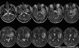
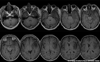
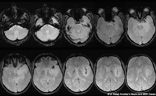
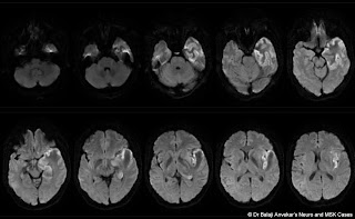
Clinical Details: middle-aged female, altered sensorium after convulsions.

TUMOR%2BMIMICS.jpg

CVT%2BMRI%2BBRAIN.jpg

CORTICAL%2BVEIN%2BTHROMBOSIS%2BMIDDLE%2B

MEDIAL%2BTEMPORAL%2BCVT.jpg

THROMBOSIS%2BPARASELLAR%2BPETROUSAL%2BSI

CVT%2BMRI%2BDIFFUSION.jpg

CVT%2BMR%2BANGIO.jpg

CVT%2BVS%2BGLIOMA.jpg


MRI study of brain shows:
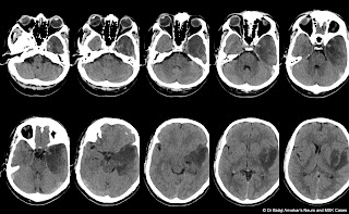
Abnormal T2 hyperintensity with marked focal parenchymal swelling due to vasogenic oedema involving left temporal, insular cortex and adjacent opercular parietal white matter. An abnormal leptomeningeal enhancement depicted on post contrast MRI along left sylvian fissure and in left medial temporal region near cavernous sinus. Multifocal low signal intensity clustered nodularity in left sylvian fissure extending towards cavernous sinus on GRE which is hyper dense on CT. No abnormal calcification on CT. Normal MR angiography of brain. No obvious aneurysm or vascular malformation on MR angio.

Left side Decompressive hemicraniectomy done.
Intraoperative findings revealed thrombosed superficial cortical veins in left sylvian fissure region and at the floor of left middle cranial fossa. 

Final diagnosis: Isolated superficial middle cerebral or Sylvian vein thrombosis.

Superficial middle cerebral vein also known as the Sylvian vein, is one of the superficial cerebral veins. It usually courses along the Sylvian fissure posteroanteriorly and drains numerous small tributaries from the opercular areas around the lateral sulcus. It curves anteriorly around the tip of the temporal lobe and drains into the sphenoparietal sinus or directly into the cavernous sinus. 

Suzuki classification of the superficial Sylvian venous drainage pathways:
1. sphenoparietal type: (54%) drains into the sphenoparietal sinus.
2. emissary type: (12%) courses along the lesser wing of sphenoid, turns inferiorly to reach the floor of the middle cranial fossa, joins the sphenoidal emissary veins, and passes through the floor to reach the pterygoid plexus.
3. cavernous type: (7%) directly drains into the anterior end of the cavernous sinus.
4. superior petrosal type: (2%) runs along the lesser wing and just before reaching the cavernous sinus, turns downward along the anterior inner wall of the middle cranial fossa, then runs along its floor medially to the foramen ovale to join the superior petrosal sinus.
5. basal type: (2%) runs along the lesser wing, turns downward along the anterior wall of the middle cranial fossa, then runs along its floor laterally to the foramen ovale over the petrous pyramid, presumably to join the transverse sinus through the lateral tentorial sinus or superior petrosal sinus.
6. squamosal type: (2%) turns directly backward along the inner aspect of the temporal squama and runs posteriorly to join the transverse sinus or lateral tentorial sinus.

View on the web

Δημοφιλείς αναρτήσεις