Κυριακή 3 Οκτωβρίου 2021

Conjunctivoductivo‐Dacryocystorhinostomy: A Novel Surgery for Intractable Canalicular Obstruction

xlomafota13 shared this article with you from Inoreader
View on the web

Novel function of fluvastatin in attenuating oxidized low-density lipoprotein-induced endothelial cell ferroptosis in a glutathione peroxidase4- and cystine-glutamate antiporter-dependent manner

xlomafota13 shared this article with you from Inoreader

Exp Ther Med. 2021 Nov;22(5):1275. doi: 10.3892/etm.2021.10710. Epub 2021 Sep 8.

ABSTRACT

Oxidized low-density lipoprotein (ox-LDL) induces endothelial cell apoptosis and dysfunction. Statins are drugs that are clinically used to lower serum cholesterol levels, and they have been shown to exert vascular protective effects. In the present study, human umbilical vein endothelial cells were transfected with scramble control siRNA or siRNA specific for glutathione peroxidase (GPx)4 or cystine-glutamate antiporter (xCT). MTT, Matrigel and Transwell assays were used to evaluate cell proliferation, tube formation and migration, respectively. The levels of TNF-α, IL-α, 4-hydroxynonenal, GPx4 and xCT expression were detected by western blot analysis. It was demonstrated that ox-LDL promoted cytokine production and reduced the proliferation, migration and angiogenesis of endothelial cells. It was also observed that ox-LDL decreased GPx4 and xC T expression and induced ferroptosis. Furthermore, the inhibition of ferroptosis by deferoxamine mesylate attenuated ox-LDL-induced endothelial cell dysfunction and restored ox-LDL-decreased GPx4 and xCT expression. Consistent with these results, GPx4 and xCT knockdown by siRNA transfection aggravated ox-LDL-induced endothelial cell dysfunction and inhibition of proliferation. To the best of our knowledge, the present study was the first to discover that fluvastatin may protect endothelial cells from ox-LDL-induced ferroptosis and dysfunction. Furthermore, knockdown of GPx4 and xCT expression blunted the protective effects of fluvastatin on ox-LDL-treated endothelial cells. These data indicated a novel function of fluvastatin in the protection of endothelial cells from ox-LDL-induced ferroptosis, the mechanism of which involves the regulation of GPx4 and xCT.

PMID:34594412 | PMC:PMC8456483 | DOI:10.3892/etm.2021.10710

View on the web

miR-122-5p downregulation attenuates lipopolysaccharide-induced acute lung injury by targeting IL1RN

xlomafota13 shared this article with you from Inoreader

Exp Ther Med. 2021 Nov;22(5):1278. doi: 10.3892/etm.2021.10713. Epub 2021 Sep 8.

ABSTRACT

MicroRNAs (miRs) and inflammatory cytokines can induce acute lung injury (ALI), which can develop into acute respiratory distress syndrome in severe cases. Previous research has revealed that miR-122-5p participates in the development of ALI, and that its expression is positively associated with ALI. However, the mechanism by which miR-122-5p contributes to ALI remains to be determined. In the current study, TargetScan and dual luciferase reporter gene assays were used to confirm that IL-1 receptor antagonist (IL1RN) was a target of miR-122-5p. Subsequently, by referring to previous literature, a lipopolysaccharide (LPS)-induced ALI cell model was established. A549 cells were transfected with mimic control or miR-122-5p mimics for 24 h, and 10 µg LPS was used to treat the transfected cells for 12 h. The results revealed that miR-122-5p mimics dec reased cell viability and promoted apoptosis. Lactate dehydrogenase (LDH) release assays indicated that miR-122-5p mimics increased LDH release. ELISA demonstrated that miR-122-5p mimics promoted TNF-α, IL-1β and IL-6 expression levels. A549 cells were transfected with inhibitor control, miR-122-5p inhibitor, miR-122-5p inhibitor + control-small interfering (si)RNA or miR-122-5p inhibitor + IL1RN-siRNA for 24 h, after which the cells were treated with 10 µg LPS for 12 h. The results revealed that the effects of the miR-122-5p inhibitor were the opposite of those of the miR-122-5p mimic. All the effects of miR-122-5p inhibitor on LPS-treated A549 cells were significantly reversed by IL1RN-siRNA. Overall, the results highlighted miR-122-5p as a potential novel target for the treatment of ALI.

PMID:34594415 | PMC:PMC8456493 | DOI:10.3892/etm.2021.10713

View on the web

Prophylactic fasciotomy of the anterolateral thigh donor site in head and neck reconstruction

xlomafota13 shared this article with you from Inoreader

1-s2.0-S0196070921X00051-cov150h.gif

Publication date: January–February 2022

Source: American Journal of Otolaryngology, Volume 43, Issue 1

Author(s): Katherine Sadaniantz, Lauren Daly, Jing Bai Qin, Jeremiah Tracy

View on the web

Medial patellar plica syndrome

xlomafota13 shared this article with you from Inoreader

Clinically young patient with anteromedial knee pain.

medial%2Bplica%2Bsyndrome.jpg


MRI shows a well-defined low signa l intensity band running across medial patellofemoral recess.
However there is no obvious associated bone marrow oedema involving medial articulating facet of patella or medial femoral trochlea. 
Associated joint effusion.
Medial plica mentioned in the report with joint effusion.

Medial plica syndrome

Synonym: synovial plicae of the knee.

Common cause of anterior knee pain typically present with pain on anteromedial aspect of the knee above the joint line associated with crepitation, catching and locking sensations. 
Typically involves young with athletic background. 
There are actually synovial invaginations as a part of remnants of embryological development. They are present in over 70% of individuals and are mostly asymptomatic.
However can become inflamed and symptomatic. They can undergo fibrosis secondary to repeate d inflammation making them non-stretchable.
In symptomatic patients, medial plica seen as low signal intensity band on T1 as well as T2-weighted images with an associated chondral defect involving medial articulating facet of patella.
Treatment is mainly conservative, physiotherapy and steroid injections.

View on the web

Morel-Lavallée lesion MRI

xlomafota13 shared this article with you from Inoreader

Clinically RTA, run over by tractor.

Morel-Lavall%25C3%25A9e%2Blesion.jpg

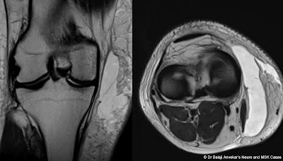
This MRI study shows a focal well defined lentiform shaped subcutaneous collection on medial aspect of knee joint superficial to the superficial fascia.
Collection is clear, hypo intense on T1-weighted images without any septation or loculation. No obvious high signal intensity methaemoglobin staining on T1-weighted images to suggest any haematoma.

Morel-Lavallée lesion

These are focal well defined lentiform shaped subcutaneous serous collections commonly encountered during MRI knee joint in the setting of severe trauma. However these lesions are typically described in thigh as well defined collection overlying the greater trochanter of the femur, tensor facia lata. 

These actually localized haemolymphatic collections secondary to closed degloving injuries, where the skin and subcutaneous fatty tissue abruptly separate from the underlying fascia owing to trauma. The potential space thus created superficial to the superficial fascia is filled by serous fluid, some times frank blood.
Similar collection secondary to similar biomechanical forces are described in lumbar region and over the scapula as well. 

The accumulated collection usually needs nothing to be done, resolves spontaneously. However may persist longer if gets organized and encapsulated. The conservative management is with compression bandages. Surgical drainage may be sufficient for larger collection. However, the capsule may need to be resected to prevent re-accumulation if it is long standing and encapsulated collection with thick organized wall. 

View on the web

Impact of Depth of Invasion (According to Layer) on Lymph Node Metastasis in Buccal Mucosa Cancers

xlomafota13 shared this article with you from Inoreader

Abstract

(1) To study layer of invasion as predictor of cervical lymph node metastasis. (2) To decide a cut off depth according to layer of invasion for neck dissection. It is a prospective study with sample size of 220 patients at tertiary care hospital with previously untreated operable buccal mucosa carcinoma over period of 21 months. The factors considered under the study were Tumor layer of invasion, lymph node metastasis, Ultrasonography doppler neck, CT scan and gender. Wide excision of the lesion and reconstruction was done. Histopathological analysis of resected specimen were recorded as part of data collection for all the cases. Quantitative data represented using Mean ± SD (Standard Deviation) and median and interquartile range compared using Chi square test. Standard principles of univariate analysis was used according to statistical methods. Depth when measured according to layer of invasion, was significantly associated with lymph no de metastasis with 89% of the muscle as layer of invasion being Lymph node positive (p = 0.009). There is 1.39 times chances of lymph nodal metastasis in patients with muscle invasion as compared to submucosal invasion. Tumour layer of invasion plays important role in predicting chances of nodal metastasis in clinically N0 neck in buccal mucosa cancer. According to the study, we should electively proceed with ipsilateral lymph node dissection once the layer of tumour invasion is muscle. This study is limited by sample size and short duration of one year and nine months.

View on the web

Δημοφιλείς αναρτήσεις